Lee J. Skandalakis Surgical Anatomy and Technique
 Sharen Selvadurai
Sign up for access to the world's latest research
AI-generated Abstract
The paper discusses the surgical anatomy and techniques associated with various procedures, with particular emphasis on the anatomy of the scalp, including its vascular, lymphatic, and nerve supplies. It presents detailed insights into the blood supply of the scalp, highlighting the important arterial branches and their anastomoses, as well as the implications for surgical procedures. Additionally, the paper outlines techniques for surgical interventions, positioning, and considerations necessary for effective outcomes.
Figures (683)


Key takeaways
- The nerve may pass anterior or posterior to the artery, or between its branches (Fig. 2.16 ).
- The ligament contains the following structures: left gastric artery and vein; hepatic division of left vagus trunk; lymph nodes; occasionally, both vagal trunks; occasionally, branches of the right gastric artery and vein; the left hepatic artery when it arises from the left gastric artery (in 23 percent of cases).
- The portal vein lies behind the pancreas and in front of the inferior vena cava, with the common bile duct on the right and the common hepatic artery on the left.
- In this ligament, the proper hepatic artery lies to the left of the common bile and hepatic ducts and anterior to the portal vein.
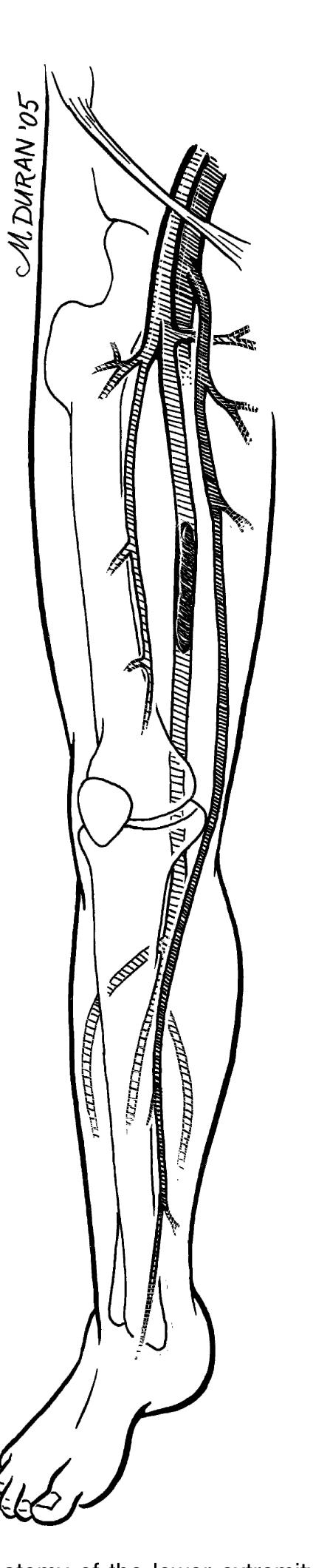
- The external iliac artery becomes the common femoral artery as it emanates from a point under the middle of the inguinal ligament.
